Neuro-anatomical tracing techniques
Objectives
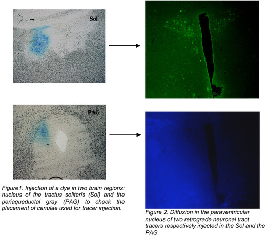
Several anterograde and retrograde neuronal tracers have been developed for respectively evidencing afferent and efferent neural pathways. These tools have largely contributed to the understanding of neuroanatomical connections between different central or peripheral organs. Recently, a new class of neuronal tract tracers consisting of modified virus, has been efficaciously developed and used (Enquist, 2005).
Summarized methodology
The general procedure consists in injecting the neuronal tracers in the organs or regions of interest. When these injections are performed in the brain, animals are placed in a stereotaxic frame and brain canulae are used for the injection of the tracers. After a post-survival period depending on the properties of the neuronal tract tracer used, animals are sacrificed and the organs of interest are collected and sliced. Then, slices are directly examined using a microscope under epifluorescence if fluorescent tracers are used (cf. confocal microscopy), or after immunocytochemical labelling if the tracers have to be detected.
Endpoints
- Qualitative analysis of the localisation of the neuronal tract tracers in organs or regions of interest
- Number and percentage of single, double or triple-labeled cells
 |
J Sex Med (2010) - Accepted

Links to applicable Targeted disorders / Pathophysiological models
- Atherosclerosis
- BPH (Benign Prostatic Hyperplasia)
- Diabetes
- ED (Erectile Dysfunction)
- Ejaculatory disorders (premature or delayed ejaculation / anejaculation)
- FSD (Female Sexual Dysfunction)
- Hypertension
- IC (Interstitial Cystitis) / Painful bladder syndrome
- Metabolic Syndrome and Obesity
- Myocardial Infarction
- NDO (Neurogenic Detrusor Overactivity)
- OAB (Overactive Bladder)
- SCI (Spinal Cord Injury)


 Download this page in PDF