


Protein expression Immunohistochemistry - Immunofluorescence
Objectives
Immunohistochemistry allows evidencing the presence a protein within a tissue section but also its localization within the tissue. This method may help to better characterize the cellular component targeted by an active compound and/or its mechanism of action.
Summarized methodology
- Immunohistochemistry can be performed on paraffin-embedded tissue sections or cryosections.
- Immunostaining can be performed with the use of an enzyme-coupled (HRP or AP) or with a biotinylated secondary antibody when signal amplification (biotin-streptavidin-HRP complex) is needed
- Immunological reaction is detected using either a colorimetric enzymatic reaction or a fluorescent dye
Endpoints
- Expression and localization of protein of interest
- semi-quantitative evaluation of the level of expression of a protein of interest (figure 1)
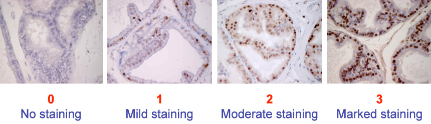 |
| Figure 1: Representative images of semi-quantitative scoring of immunostaining (PCNA) in rat prostate sections depending on intensity of the staining (pelvipharm internal data). |
Related Pelvipharm bibliography
Gelez, H et al.
J Sex Med (2010) : 7 (suppl 4):151 (ISSM 2010)
J Sex Med (2010) : 7 (suppl 4):151 (ISSM 2010)

Links to applicable Targeted disorders / Pathophysiological models
- Atherosclerosis
- BPH (Benign Prostatic Hyperplasia)
- Diabetes
- ED (Erectile Dysfunction)
- Ejaculatory disorders (premature or delayed ejaculation / anejaculation)
- FSD (Female Sexual Dysfunction)
- Hypertension
- IC (Interstitial Cystitis) / Painful bladder syndrome
- Metabolic Syndrome and Obesity
- Myocardial Infarction
- NDO (Neurogenic Detrusor Overactivity)
- OAB (Overactive Bladder)
- SCI (Spinal Cord Injury)
 Download this page in PDF